Supplier:
Aviva Systems Biology IncorporatedMouse Anti- Bcl- 2- UNLB(0.1 mg)
Prices direct from Aviva Systems Biology Incorporated
Quick response times
Exclusive Absave savings/discounts
SPECIFICATIONS
Size
0.1 mg
Hosts
Mouse
Clone
10C2
Format
Purified (UNLB) Antibody
Presku
OASB01185
Isotype
IgG1
SUPPLIER INFO


Latest promotions
Buy any polyclonal or monoclonal antibody from our extensive range of pre-made antibodies and for a limited time only receive a $50 discount!(T&C apply:...
New brilliant antibodies, and new lower prices!For flow cytometry reagents in general, \"bright is better.\" The violet-excitable BD Horizon™ BV421 and...
Did your supplier increase the price of Fetal Bovine Serum? Did they substitute the US Origin with USDA? Well say no more! Innovative Research is still...
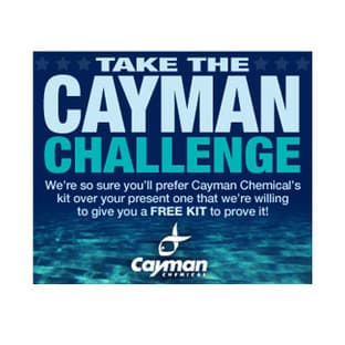
We're so sure that you'll prefer Cayman Assay kits over your present brand that we're willing to give you a free assay kit to prove it!

For the past decade scientists have extensively used ATS secondary toxin conjugates to make their own targeted toxins for in vitro use.The ability to combine...
10% Discount on 2 Rabbit Polyclonal Antibody Service. With over 20 years experience, SDIX has developed into the premier US custom antibody producer,...
Bulk Cytokines with Custom Vialing.20 - 50% off cytokines, growth factors, chemokines and more...For a limited time Cell Sciences is offering substantial...
Are you planning to have a customised antibody made for your research?Since 2000, Everest has been producing a catalog containing thousands of affinity...
Top suppliers

Agrisera AB
11 products
Biotrend
Biosensis
969 products
ABBIOTEC
3011 products
SDIX
1 products
Spring Bioscience
2291 products
Cell Signaling Technology
4976 products
Rockland Immunochemicals, Inc.
7592 products
Boster Immunoleader
1533 products
OriGene Technologies Inc.
5281 products
Maine Biotechnology Services
227 products

BD (Becton, Dickinson and Company)
1 products
ABNOVA CORPORATION
Randox Life Sciences
1502 products